
MAP and Johne’s Disease in General
 Mycobacterium avium subspecies paratuberculosis, or MAP, is a small (0.5 x 1.5 microns) rod-shaped bacterium that has a rough waxy cell wall with a trilaminar structure. This wall is composed of a thick waxy mixture of unique lipids and polysaccharides but lacks glycolipid antigens on its surface. This kind of cell wall facilitates the mycobacterium’s resistance to physical factor’s (e.g. heat, cold, sunlight, drying. etc.) and common disinfectants like chlorine. If MAP is found in soil or water samples, it can survive (but not grow and multiply) for over a year after fecal contamination via an infected animal.
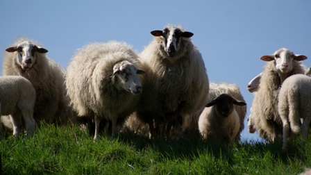 MAP was first identified as a pathogen in the late 1800s, when it was determined to be the cause of Johne’s (pronounced YO-nees) disease in cattle. Johne’s disease, sometimes called paratuberculosis or Bovine Johne’s Disease (BJD), is a chronic, fatal, gastrointestinal wasting disease that primarily affects the small intestine of ruminants like cattle, deer, sheep, goats, antelope, bison and even camels! Both domestic and wild animals can contract Johne’s disease. Although other animals occasionally become MAP-infected it does not usually result in disease.
How MAP Causes Disease on the Farm
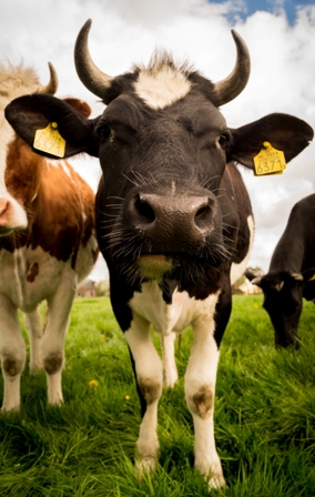 Johne’s disease is most common in dairy cows. This is likely due to frequent exposure to the fecal matter of an infected animal, common in the conditions in which dairy herds are raised. MAP is shed in the fecal matter of animals, even if they are not showing signs of disease. In fact, primary exposure to MAP generally begins at birth where the bacteria is passed to calves via milk or from fecal matter on the mother’s teats. Despite this early infection, MAP cannot be detected until the cow is two years old, long after they’ve been shedding the pathogen into common areas on the farm. A cow will only show signs of infection when the disease is at an advanced stage, which makes MAP very difficult to eradicate from the herd. Although contact with fecal matter is the primary exposure route, MAP can be passed from one animal to another via milk, soil and water.
 One interesting 2009 study examines how MAP is able to survive on different types of water troughs in biofilms. (Biofilm is essentially the slimy substance produced by bacteria grouping together in a wet environment and forming a protective outside layer. It’s the ring around the drain in your sink, the plaque on your teeth and the slippery substance on rocks in a stream.) Partially due to its production of biofilm, MAP can survive for up to a year in the environment due to its resistance to heat, cold and drying. It’s one tough bug!
Physical and Histological Symptoms of Johne’s Disease
The symptoms of Johne’s disease are eerily similar to those of Crohn’s disease. An infected cow will initially show no signs of disease or distress. As the disease progresses, after a 3-5 year incubation period, the infected cow starts rapidly losing weight loss and develops diarrhea. Despite continuing to eat, the cow becomes emaciated and weak. A fever is not common at this late stage of disease. In dairy cows, milk production often decreases. Eventually the animal will starve, despite having a good appetite and eating a healthy diet.
The histological markers (or how the tissue looks at a microscopic level) of Johne’s disease explain this starvation. When MAP is ingested by the animal, the bacteria are taken up by specialized cells in the part of the small intestine called the ileum, where nutrients are absorbed from feed. The wall of the ileum contains a large number of pockets of lymphoid tissue known as Peyer’s patches that lie just beneath the interior surface of the intestine. Peyer’s patches are clusters of macrophages and lymphocytes (white blood cells) that are organized much like lymph nodes. Covering Peyer’s patches are a layer of cells called M cells. These cells function to circulate into the lumen of the intestines where they ingest antigens (bacteria) before returning to the Peyer’s patch to “show” these antigens to the macrophages and lymphocytes. This is a means of “educating” the cells in a young animal about its environment and is a protective mechanism designed to help the animal become immune to pathogens in its environment.
Unfortunately, when M cells bring MAP to the Peyer’s patch, the bacteria finds an ideal place for growth. Macrophages in Peyer’s patches engulf MAP with the intention of destroying the foreign invader, but MAP out-smarts the immune system and starts slowly multiplying inside these cells. MAP multiplication continues until it eventually kills the cell, spreads and infects other nearby cells. In time, other parts of the ileum and other regions of the body are teaming with millions of the mycobacteria.
As the animal’s body tries to rid itself of these bacteria, the immune response causes a characteristic inflammation pattern, called granulomatous inflammation. Eventually, this type of inflammation thickens the intestinal lining, preventing it from functioning normally. This leads to poor absorption of nutrients and eventual diarrhea. As a result, although animals may be feeling and eating well, they begin to lose weight gradually until they succumb to the disease.
 MAP in cows is highly infectious via milk, water and manure and generally fatal. MAP is thus a slow, chronic, progressive infection using strategies for infection very similar to those of the more well-known bacterial infection tuberculosis (TB). In all of the species studied, it’s the very young that are the most susceptible to MAP. Animals generally develop some measure of protection against MAP over time as the immune system matures. Peyer’s patches are most active and numerous in the very young, and these may provide an explanation for the high susceptibility of neonates and the initial site of infection, the ileum. At some point later in life, MAP starts to replicate and when the immune system responds, granulomatous inflammation causes a cascade of disease progression.
Prevalence of Johne’s Disease in the World
 Today, Johne’s disease has been found in almost every country that has tested their animals. It’s estimated that 91% of United States dairy herds have at least one animal with Johne’s disease. In comparison, only about 7-8% of beef herds are infected. The percentages are similar throughout the world. Each year, Johne’s disease costs the United States dairy industry between $200-$250 million. Some countries are making progress in reducing the prevalence of Johne’s disease. Australia is one country implementing measures to stop the spread of Johne’s disease.
Preventing and Reducing the Spread of Johne’s Disease
The best way to prevent Johne’s disease is to keep infected animals out of the herd. Farmers should only purchase animals from test-negative herds. For infected herds, infection control requires good herd management and regular testing. Calves should be born in a clean environment and their exposure to manure from adult animals should be minimized. Avoiding manure contamination in other areas, like feed and water sources is important. Identify and remove, or keep separate all test positive animals.
The three basic ways to test an animal for Johne’s disease are (1) by obtaining a fecal sample and testing for MAP by culturing it (a process taking 2-3 months) or by finding MAP DNA in the sample using a technology called PCR, or (2) testing the blood for antibodies to MAP in blood or milk samples.
 Symposium presenter Dr. Michael T. Collins is one of the world’s foremost experts on Johne’s disease. His site, the Johne’s Disease Information Center, provides detailed information and resources for those wanting more information about Johne’s disease. The Paratuberculosis Awareness and Research Association also gives detailed information and up to date information surrounding this subject.
Human Exposure to MAP
While MAP is highly contagious within the cattle population, the same principles of bacterial transmission do not hold true when applied to humans. Being a non-ruminant, the human is an unwilling or abnormal host for MAP. It’s like trying to fit a square peg in a round hole. MAP thrives in the higher body temperature of cattle (101.5 degrees Fahrenheit) and needs to adapt to the human environment to survive. It needs to “shed some skin.” How it does that is ingenious, and discussed in detail in the Crohn’s Info section. So while MAP is not transmitted to humans the same way as it is between cattle, the risk of human exposure remains a threat.
 The belief that exposure to MAP is reserved for people who work on dairy farms is wholly incorrect. In fact, most people are exposed by drinking pasteurized milk. If the cow’s teats are contaminated with MAP during milking (a common occurrence) the bacteria will contaminate the milk. While pasteurization kills most bacteria, it does not reliably kill all MAP. From reliable testing, about 2% of retail milk samples contain viable (live) MAP bacteria. Even more frightening is that MAP is prevalent in infant formula. Using the best technology available, 60 infant formula samples were tested for the presence of live MAP bacteria. Overall, 40% were positive and a staggering 56% of the samples from the United States were positive. Based on estimates of how many MAP bacteria survive in milk and formula, a child might consume between 1,000 and 100,000 MAP bacteria by the age of ten. Keep in mind, as stated above, it is the very young animals in the herd that are most susceptible to contracting an infection.
 Another distasteful process common in herds with Johne’s disease is culling. Animals that test positive or show signs of disease are removed and sold to the beef market to be used for ground beef or processed meat. Even cooking to a well done temperature may not kill all of the MAP bacteria in the meat. While it’s admirable that sick animals are removed from the dairy supply chain, it’s counterproductive to then put them in the beef supply chain. Recall that animals with Johne’s disease do not always show disease symptoms.
What do MAP and Johne’s disease have to do with Crohn’s Disease?
The connection between MAP and Crohn’s disease was first studied over 100 years ago. In 1913, Dalziel noted similarities in the intestines of sick humans and cattle suffering from Johne’s disease. He proposed that Johne’s disease and what would become Crohn’s disease were the same. Fast forward to 1932 and the discovery of Crohn’s disease. Strikingly similar symptoms and the characteristic granulomatous inflammation associated with Johne’s disease began to show up in increasing numbers in the human population.
As the human diet has shifted to include more meat and milk, the incidence of Crohn’s disease has increased. This correlation does not prove causation, however, new research from John Aitken’s lab has identified a close relative of MAP in many Crohn’s disease patients. When Crohn’s disease patients are treated with an antibiotic regimen that seeks to eradicate MAP, remission is achieved at a greater rate than that of Remicade or Humira. However, until MAP is declared a human pathogen by the FDA, the USDA or other world governments, no control measures will be mandated to stop the spread of MAP to the human population. Read more about the treatment of Crohn’s disease with Anti-MAP therapy in the Crohn’s Info section.
For a more thorough understanding of MAP, Johne’s disease and the link to Crohn’s disease, watch Dr. Collins’ illustrated video or his symposium presentation.